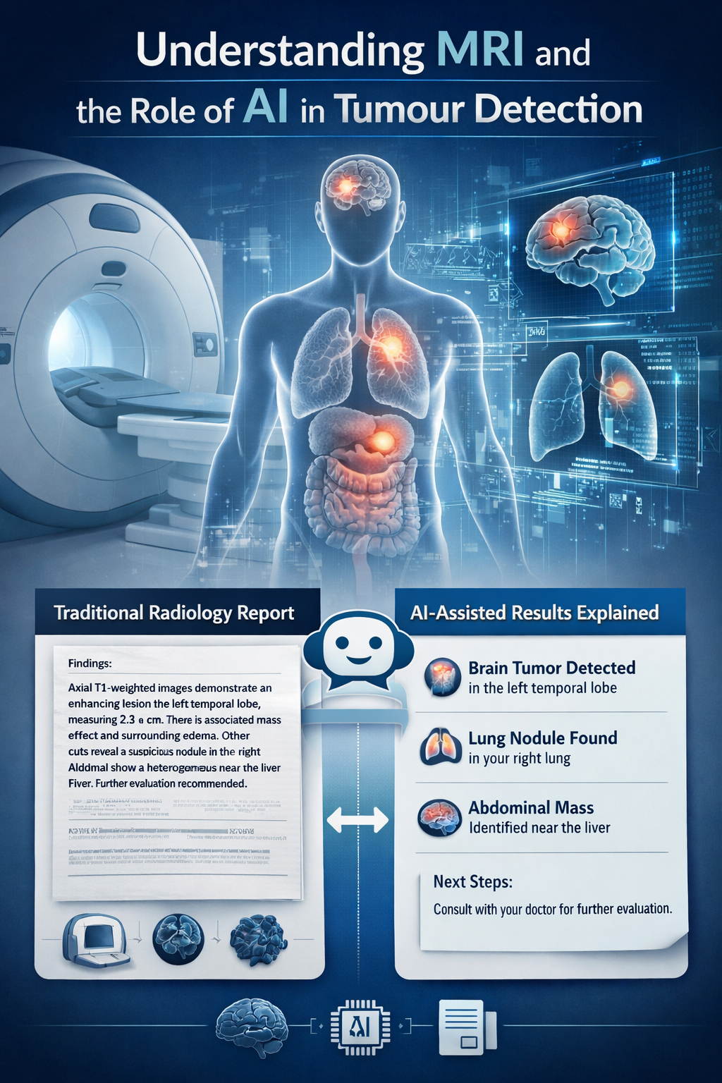
Understanding MRI and Its Role in Tumour Detection
MRI is one of those medical terms everyone has heard, often during a hospital visit or in a movie scene, yet few people truly understand how it works. Magnetic Resonance Imaging (MRI) is one of the most advanced and widely used tools in modern medicine. It uses a powerful magnetic field and radio waves to align hydrogen atoms in the body and detect the signals they emit as they return to their normal state. These signals are then used to create highly detailed images of internal organs and tissues such as the brain, liver, and muscles. Unlike X rays or CT scans, MRI does not use radiation.
In the context of tumour detection, MRI plays a crucial role. It helps radiologists and oncologists identify the exact location, size, and nature of a tumour, as well as determine whether it is spreading to nearby tissues. MRI scans allow radiologists to describe the tumour’s shape, margins, blood supply, and its relationship with surrounding organs, all of which are essential for diagnosis and treatment planning, including surgery or radiation therapy.
After the scan, radiologists prepare a detailed report and provide their opinion on the findings. However, these reports are often written in highly technical medical language filled with jargon such as “heterogeneous enhancement,” “lesion margins,” or “contrast enhancement.” For most patients, understanding such reports without a doctor’s explanation is almost impossible. This is where chatbots like ChatGPT and Deepseek are beginning to make a difference.
What Are Chatbots and How They Are Trained to Read MRI Reports
Chatbots are artificial intelligence programs designed to understand and respond to human conversation. Modern chatbots are powered by generative AI, a form of advanced artificial intelligence that can create new text, ideas, or responses based on the data it has learned. Instead of following fixed commands, generative AI understands context, reasons through information, and produces human like responses in real time.
When chatbots are built using large language models, they can process vast amounts of data, including complex scientific and medical information. These models are trained on millions of examples from medical textbooks, research journals, clinical guidelines, and radiology reports. This training enables them to understand how doctors describe diseases and interpret imaging findings. Over time, the chatbot learns to recognize patterns, interpret medical terminology, and explain complex concepts in simple, patient friendly language.
In medical imaging, AI chatbots such as RadiologyGPT, Glass AI, Qure.ai Assistant, and Tempus, formerly known as Arterys, are increasingly being used to assist radiologists. These tools help interpret scans, generate structured reports, and detect abnormalities. RadiologyGPT and Glass AI are designed to understand radiology specific terminology and summarize imaging findings. Qure.ai Assistant, developed in India, is widely used for analyzing chest X rays, CT scans, and brain images to identify early signs of disease. Tempus integrates AI driven imaging insights with clinical and genomic data to support precision medicine. Together, these technologies are transforming radiology by improving efficiency, accuracy, and accessibility in diagnostic imaging.
The Research Behind AI Chatbots in MRI Interpretation
The research was conducted at Peking Union Medical College Hospital, Chinese Academy of Medical Sciences, Beijing, China, and was published in the journal Scientific Reports on 25 August 2025. It was a cross sectional analysis involving 6,174 MRI reports from tumour patients who underwent scans between January 1, 2019, and December 31, 2024, across three hospitals. Two artificial intelligence chatbots, GPT o1 preview (Chatbot 1) and Deepseek R1 (Chatbot 2), were evaluated for their ability to interpret MRI reports, classify tumour characteristics, assess the need for surgery, and suggest possible treatment options.
The MRI reports, originally interpreted by radiologists, included detailed descriptions of normal anatomical structures and abnormal lesion signals, along with preliminary diagnoses. Two independent reviewers examined the original MRI reports and corresponding images, categorizing the findings as benign, atypical, or malignant. In cases of disagreement, a third oncologist made the final decision. To ensure patient confidentiality, all identifying information such as patient names, registration numbers, examination dates, and physician details was anonymized before analysis.
The primary objective of the study was to evaluate the performance of AI chatbots like ChatGPT and Deepseek R1 in translating complex MRI reports into patient friendly language while also offering clinical recommendations. The chatbots were asked to answer four specific questions based on each report. First, to explain the report in terms understandable to a layperson. Second, to classify the lesion as benign, atypical, or malignant. Third, to determine whether surgery was required. Fourth, to recommend an appropriate treatment plan based on the findings.
All chatbot generated responses were reviewed by medical experts. Each response was independently evaluated by two medical reviewers, and disagreements were resolved by consulting a third oncologist to ensure accuracy and fairness.
Key Findings from the Research
The results showed that both AI chatbots significantly improved the readability and accessibility of MRI reports compared to those written by radiologists. However, Deepseek R1 consistently outperformed GPT o1 preview across several parameters, including accuracy, readability, empathy, and clinical relevance.
The original MRI reports were highly technical and difficult for patients and their families to understand. When interpreted by the chatbots, the language became much simpler and more accessible. In terms of medical accuracy, Deepseek R1 again performed better. It correctly classified tumours as benign, atypical, or malignant in 92.05 percent of cases, compared to 89.03 percent for GPT o1 preview. For assessing the need for surgery, Deepseek R1 achieved an accuracy of 95.12 percent, while GPT o1 preview achieved 84.73 percent.
The study also evaluated empathy and clinical usefulness. Both chatbots demonstrated a high level of empathy in their responses, with median empathy scores of around 4 on a 5 point Likert scale.
However, the results were not without limitations. Some errors occurred due to what are known as hallucinations, a common issue with AI chatbots where incorrect or fabricated information is generated. In this study, hallucinations led to misinterpretation of certain medical terms such as “heterogeneous enhancement” or “invasion,” resulting in incorrect tumour classification and inaccurate assessments. These findings highlight that AI chatbots are still far from perfect when dealing with complex medical language.
The Future Role of AI Chatbots in Medical Imaging
This study highlights a promising future for AI chatbots in medical imaging, including sonography, X ray, CT scans, and MRI. These tools have the potential to transform how imaging information is communicated by making reports more understandable for patients, providing instant explanations, and reducing the workload of doctors who spend considerable time simplifying complex findings.
In my opinion, the future of healthcare will inevitably move toward AI assisted practice. Hospitals will increasingly adopt systems where doctors and AI tools work together. Rather than replacing medical professionals, AI should be viewed as a supportive tool that provides a second opinion and helps ensure that important findings are not missed due to human error. Such collaboration can improve diagnostic accuracy and enhance the overall quality of patient care.
That said, complete replacement of doctors by chatbots is neither possible nor advisable. Medical supervision remains essential to verify AI interpretations. Ethical considerations and data privacy will also play a crucial role as AI becomes more integrated into clinical practice. AI should be seen as complementary rather than competitive. While it cannot replace the human touch in medicine, it will undoubtedly become one of the doctor’s most powerful allies.
A community based study in Ahmedabad found 33.5% antibiotic use, with over one third obtained without prescription. High consumption of ‘Watch’ group drugs and misuse among children raise concerns about antimicrobial resistance, highlighting the urgent need for rational prescribing and public awareness.
Chronic friction from tightly worn sarees and dhotis can cause waist dermatoses that rarely progress to squamous cell carcinoma. A systematic review highlights risk factors like poor hygiene and delayed care. Early detection and simple preventive measures can effectively reduce this avoidable skin cancer risk.
A cross sectional study in India reveals gaps in data driven decision making among sub district public health administrators. Despite data availability, limited training, infrastructure, and time constraints hinder effective use. Strengthening data literacy and integrating data into routine workflows is essential for improving public health outcomes.